Can we Adjust Focus to Enhance Optical Imaging Signals?
Optical imaging of intrinsic signals (OIS) has been around for almost 40 years. It is part of the basic portfolio of technologies available to neuroscientists today. This versatile technique measures small changes in light absorption that occur in the brain tissue to assess its function. Adjusting focus to enhance optical imaging signals can significantly improve the accuracy of these measurements. The signals measured in OIS are primarily related to changes in blood volume and oxygenation of the brain tissue.
One of the basic principles behind the use of OIS stems from the fact that hemodynamic changes can be used as a proxy of the neuronal activity. In brief, when neurons become active, they consume more oxygen, leading to localized changes in hemoglobin oxygenation levels and increased blood flow. Some of these variations are very small (in the order of 0.1% to 1%) and are due to changes in the absorption of the incident light by the hemoglobin molecule. Oxygenated and deoxygenated hemoglobin have distinct absorption levels at different illumination wavelengths in the visible spectrum. OIS takes advantage from these spectral properties to estimate the different contributions of the local oxygen variation and blood volume to the hemodynamic response.
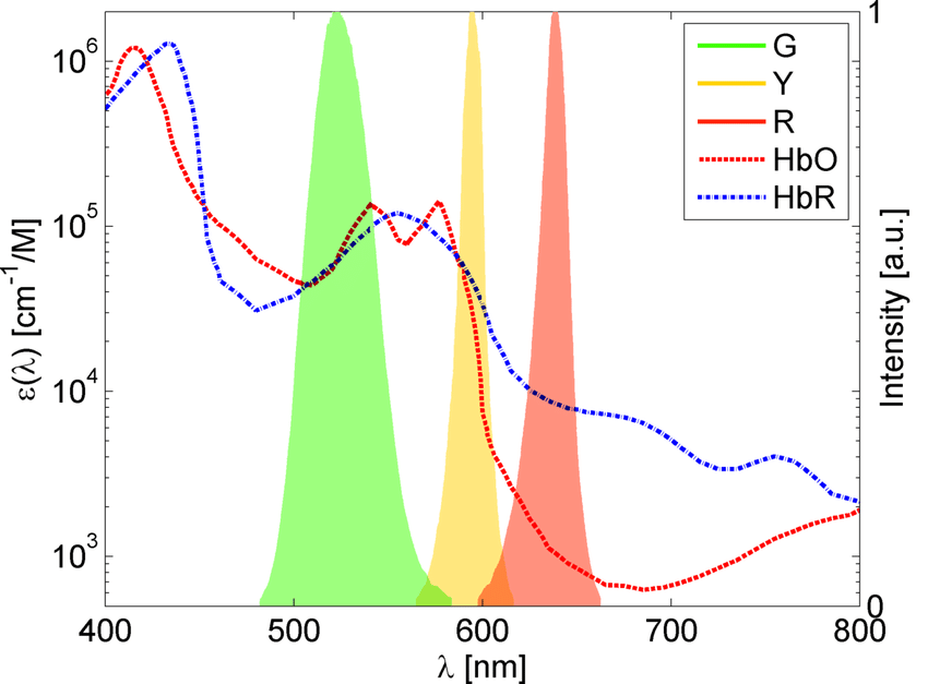
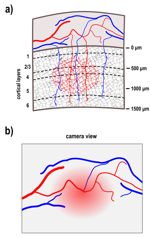
It is well known that the brain tissue scatters light differently as a function of the wavelength. In general, shorter wavelengths scatter more and than longer wavelengths which means that longer wavelengths penetrate deeper in the tissue. Different studies have estimated the extent of brain volume that contribute to the hemodynamic signal under different wavelengths (480 nm to 630 nm) and concluded that most of the detected signal comes from depths up to ~1 mm with the more superficial tissue (~500 microns) contributing the most to the hemodynamic signal composition and deeper sections contributing progressively less. For instance, the mouse cortex is about ~1.5 mm deep with a vasculature comprised of superficial arteries and veins with branches that plunge into the cortex where a web of capillaries is present. So, based on the abovementioned properties of the tissue scattering, the intrinsic signal of the mouse cortex is mostly comprised of the hemodynamic signals of superficial blood vessels followed by the signals from capillaries up to layer V.
Due to its well-localized signal, the hemodynamic responses of capillaries have been utilized by numerous researchers interested in cortical mapping. In this context, the signals from superficial blood vessels may be somewhat undesirable. Given that the spectral properties of brain tissue are fixed, what strategies can be employed to attenuate the signals from superficial blood vessels?
To address this question, we evaluated the impact of focus depth and depth of field (DOF) on the quality of recorded intrinsic signals. This aims to provide guidance on adjusting these parameters to enhance the quality of cortical maps using optical imaging of intrinsic signals.
The Experiment
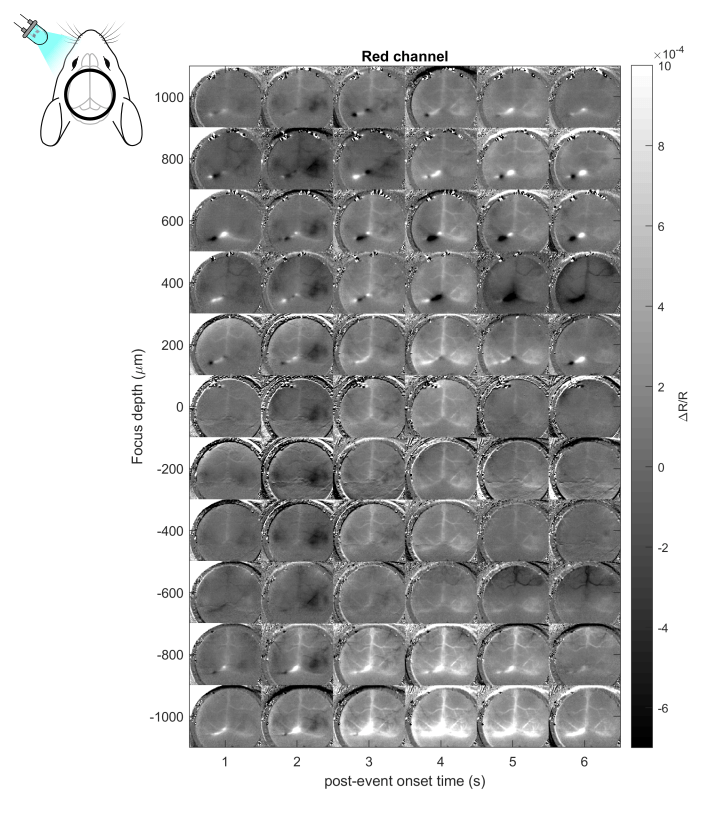
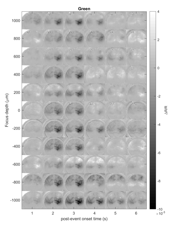
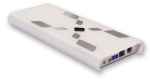
Our experiment was performed in wild-type mice under urethane anesthesia. In brief, cortical responses in the right visual cortex were elicited using a flash stimulus on the contralateral eye. The visual stimulus was repeated 30 times, and the average cortical activation was calculated to get the DeltaR/R. We recorded the cortical responses under 525nm (green) and 625 nm (red) illumination at 1000 microns above and below the cortical surface (zero focus depth set under green illumination with surface vessels in focus) spaced at 200 microns at f/1.2 using our OiS200 LightTrack Imaging device. For the depth-of-focus test, we compared 3 fStops (1.2, 2 and 2.8) of the camera lens. Our observations are summarized below.
Focus Depth
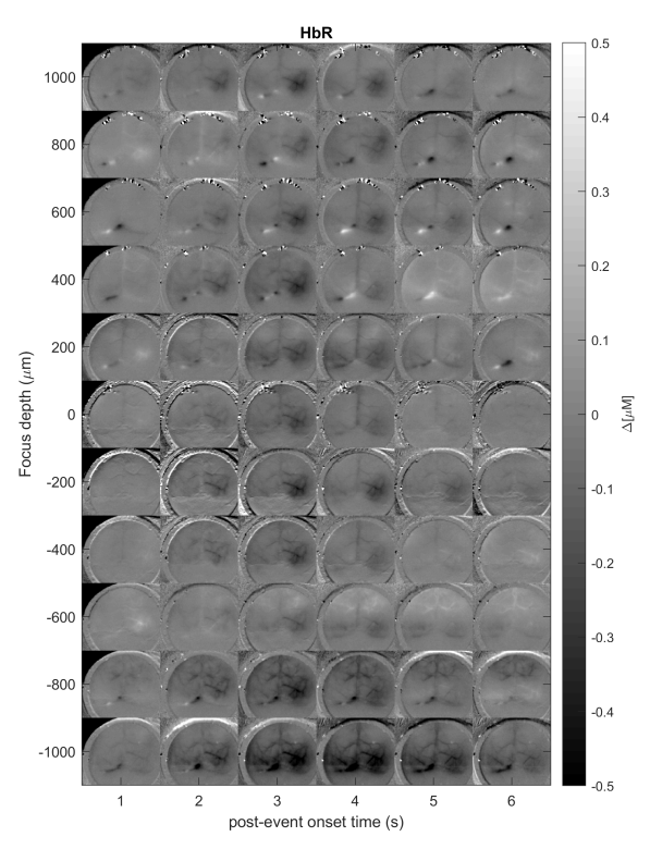
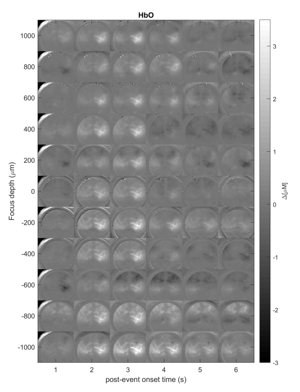
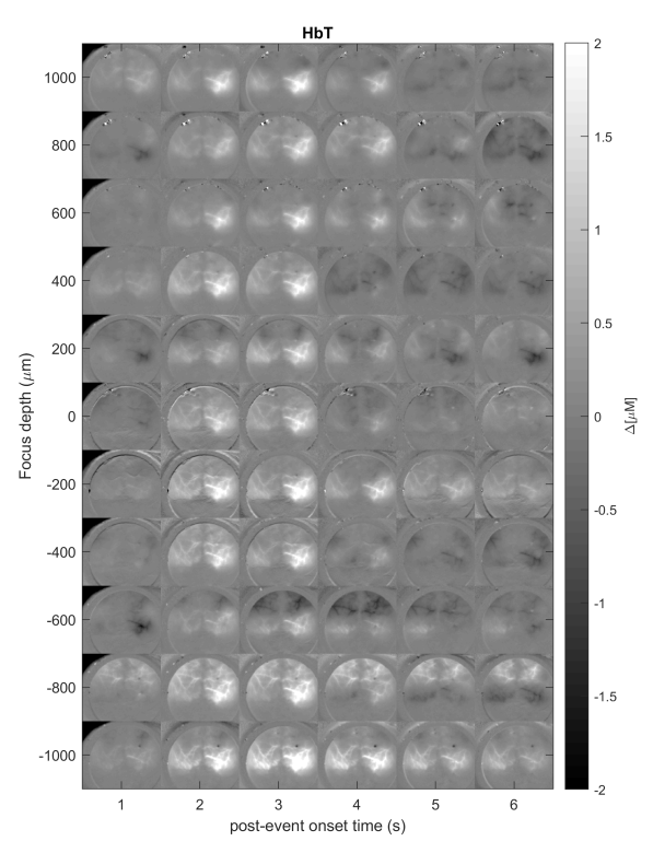
All recordings showed a local activation of the visual cortex characterized by a negative deflection of the signal. This localized activation peaked around 2 seconds after the visual stimulus onset and was followed by a positive deflection of the hemodynamic signal, now mostly confined to venules and larger veins. Although there was not a lot of change in the profile of the local activation, changing the focus depth mostly impacted the visibility of the signals from the surface blood vessels where they were reduced in a narrow band between 200 and 400 microns below the cortical surface.
Below, are the cortical responses as a function of focus depth in the red channel (left) as well as in the green channel and for the oxygenated (HbO), deoxygenated (HbR) and total hemoglobin (HbT):
Depth of Field (DOF)
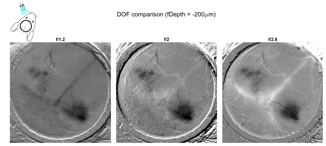
In this part of the experiment, we aimed to assess the impact of the DOF on the intrinsic signal. Instead of varying the focus plane, in this case, we increased the amount of cortex volume in focus by increasing the fStop value at the system’s lens. We observed that f/2.0 and f/2.8 made the surface blood vessels (arteries and veins) more distinguishable. The local activation was not significantly impacted by the changes in the depth-of-focus:
Adjusting Focus Enhances Optical Imaging Signals
This experiment shows that, by adjusting focus, we were able to enhance the quality of optical imaging signals. The primary impact of adjusting focus depth and depth of field on signal quality is predominantly associated with the visibility of superficial blood vessels. While this may not inherently pose a problem, it is crucial for researchers seeking to filter out these signals to consider optimizing these parameters. Based on our experiment, signal quality can be optimized by focusing slightly deeper into the cortex (approximately 300 microns) and employing a shallow depth of field (f/1.2). This approach will attenuate the signals from superficial blood vessels, thereby enhancing the detection of localized neuronal activation. Some trial and error may be necessary to achieve the optimal focus depth and depth of field.
Products Used

Don’t miss out! Subscribe to our newsletter for the latest blog updates.
We hope you found this post helpful! If you have any questions or need further information, feel free to reach out. We’re here to help!