Cortical Dynamics in Mouse Behavior Using Widefield Calcium Imaging – Part Two
In the previous blog, we explored the dynamics of cortical activity during distinct behaviors in an awake mouse. In this part, we will showcase another analysis that assesses the interconnectivity of cortical regions.
Cortical Functional Connectivity (FC)
As we mentioned in the previous blog, the mouse cortex is a complex structure with distinct modules that are functionally interconnected. This interconnection is mainly characterized by the co-activation of different brain regions over time, known as functional connectivity. The functional connectivity analysis is defined as the temporal correlation between the neuronal activities of these areas (van den Heuvel and Hulshoff Pol 2010). Several studies have characterized distinct patterns of co-activation between brain regions linked to different behavioral states. These network patterns provide insights into how the brain processes information and how changes in these patterns during pathological states reveal important clues about the underlying mechanisms of various neurological disorders such as Alzheimer’s (Dennis and Thompson 2014) and depression (Xu et al. 2024).
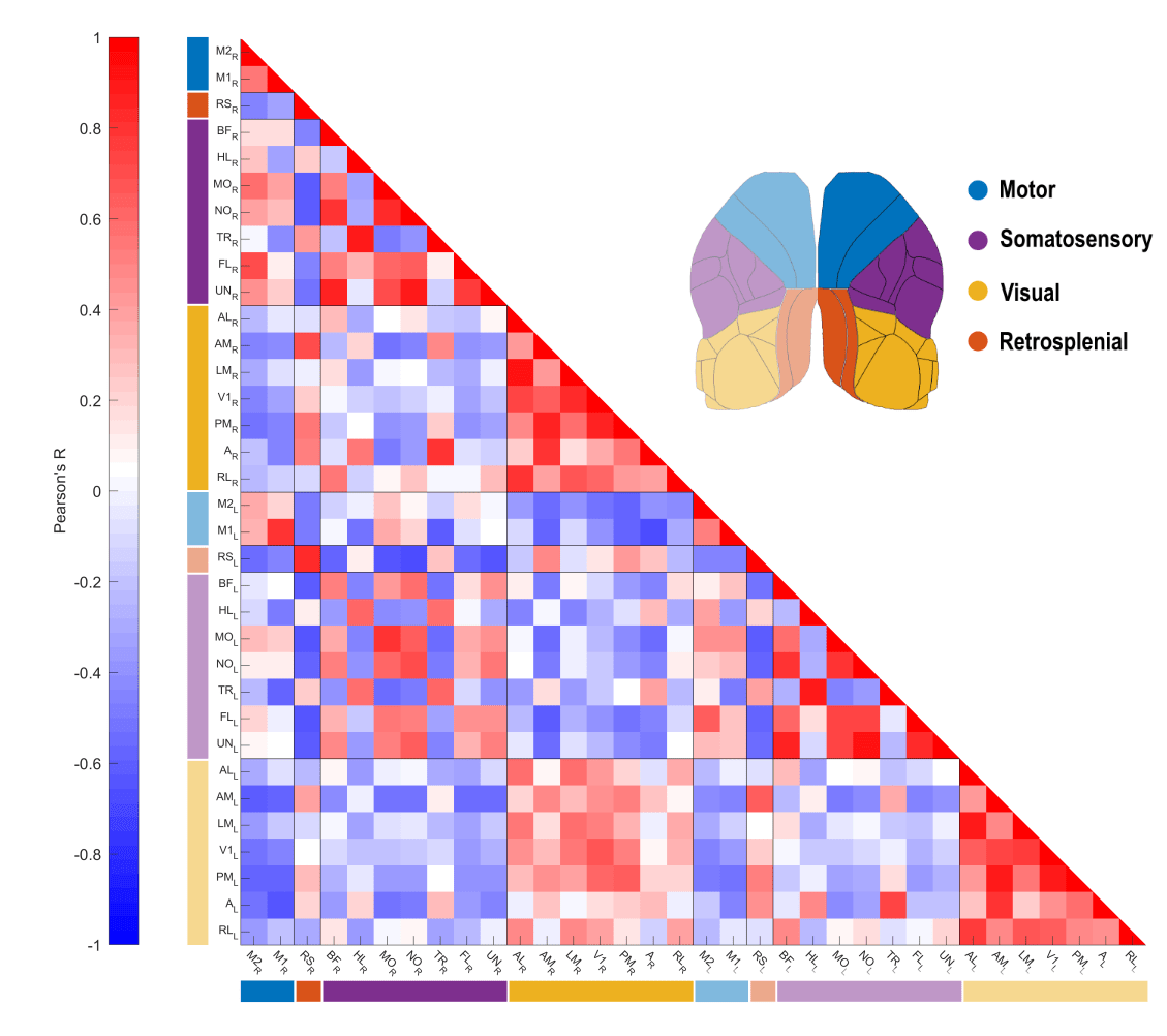
Resting state functional connectivity of a mouse cortex using the seed-pixel correlation technique.
The Resting State FC
In this part of the blog series, we continue with the data exploration of the different behavioral states classified in the previous analysis. Here, we performed the resting state functional connectivity analysis. As the name implies, resting state FC assesses the cortical interconnectivity while the animal is at rest. The anatomy and size of the mouse neocortex make it an ideal animal model to study the cortical FC using widefield optical imaging. Indeed, this approach has been widely used in widefield imaging in mice to characterize the cortical network function and how it is impacted by brain damage such as hypoxia (Bakker et al. 2023) or stroke (Bauer et al. 2014; Balbi et al. 2019), for instance.
The FC Analysis
In our first analysis, we used the behavioral camera and the treadmill data to identify and classify the animal’s movements. In order to assess the resting state FC, we removed the global oscillation from the fluorescence signal (click here for more info) and performed the temporal correlation between brain regions during moments where the animal was at rest (i.e., periods in between the behavioral states). For each resting period, we calculated the temporal correlation between the average pixel values of the cortical areas and Pearson’s R value averaged. A correlation matrix was generated, showing the functional connectivity between cortical regions.
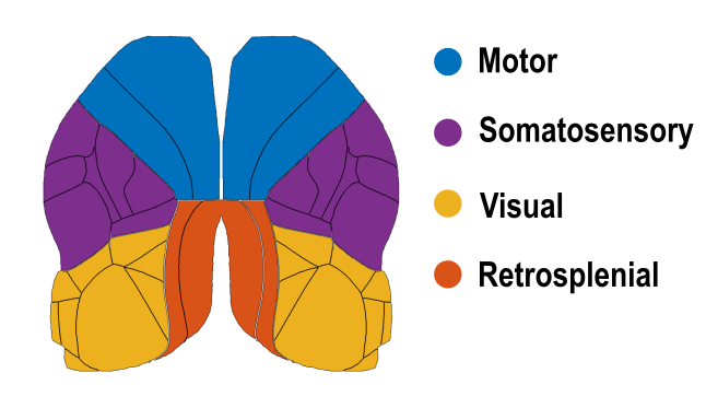
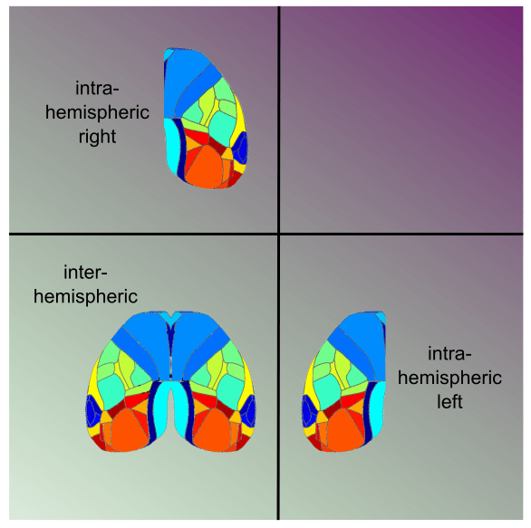
The correlation matrix is a good and straightforward way to visualize the FC between regions. The matrix is organized so the areas are clustered by functional module and hemisphere so the intra and inter-hemispheric connections are easily identified.
Below is the correlation matrix showing the resting state functional connectivity between cortical regions from the motor, retrosplenial, somatosensory and visual areas. This mouse shows a typical resting state FC profile where we can see stronger intra-hemispheric correlations within functional modules and less pronounced correlation values for inter-hemispheric connections. Regarding the connectivity between functional modules, motor regions are more strongly connected to the somatosensory areas while the visual areas exhibit higher correlations with the retrosplenial cortex. On the other hand, visual areas seem to be anti-correlated with motor areas and are weakly correlated with somatosensory areas.
Summary and Insights
In this two-part blog series, we demonstrated different ways of exploring the cortical dynamics during behavior using a very simple experimental setup. By recording an awake, behaving mouse, we were able to identify cortical patterns associated with specific behaviors. In this blog, we also showed how to assess the cortical functional connectivity during the periods between movements. This experiment highlights the strengths of the widefield optical imaging technique in mice and its great potential to help researchers better understand the underlying neuronal mechanisms of brain function.
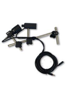
Products Used


References
Bakker, Marleen E., Ismaël Djerourou, Samuel Belanger, Frédéric Lesage, and Matthieu P. Vanni. 2023. “Alteration of Functional Connectivity despite Preserved Cerebral Oxygenation during Acute Hypoxia.” Scientific Reports 13 (1): 13269. https://doi.org/10.1038/s41598-023-40321-3.
Balbi, Matilde, Matthieu P Vanni, Max J Vega, Gergely Silasi, Yuki Sekino, Jamie D Boyd, Jeffrey M LeDue, and Timothy H Murphy. 2019. “Longitudinal Monitoring of Mesoscopic Cortical Activity in a Mouse Model of Microinfarcts Reveals Dissociations with Behavioral and Motor Function.” Journal of Cerebral Blood Flow & Metabolism 39 (8): 1486–1500. https://doi.org/10.1177/0271678X18763428.
Bauer, Adam Q., Andrew W. Kraft, Patrick W. Wright, Abraham Z. Snyder, Jin-Moo Lee, and Joseph P. Culver. 2014. “Optical Imaging of Disrupted Functional Connectivity Following Ischemic Stroke in Mice.” NeuroImage 99 (October):388–401. https://doi.org/10.1016/j.neuroimage.2014.05.051.
Dennis, Emily L., and Paul M. Thompson. 2014. “Functional Brain Connectivity Using fMRI in Aging and Alzheimer’s Disease.” Neuropsychology Review 24 (1): 49–62. https://doi.org/10.1007/s11065-014-9249-6.
Heuvel, Martijn P. van den, and Hilleke E. Hulshoff Pol. 2010. “Exploring the Brain Network: A Review on Resting-State fMRI Functional Connectivity.” European Neuropsychopharmacology 20 (8): 519–34. https://doi.org/10.1016/j.euroneuro.2010.03.008.
Xu, Ming, Xuemei Li, Teng Teng, Yang Huang, Mengqi Liu, Yicheng Long, Fajin Lv, et al. 2024. “Reconfiguration of Structural and Functional Connectivity Coupling in Patient Subgroups With Adolescent Depression.” JAMA Network Open 7 (3): e241933. https://doi.org/10.1001/jamanetworkopen.2024.1933.
Don’t miss out! Subscribe to our newsletter for the latest blog updates.
We hope you found this post helpful! If you have any questions or need further information, feel free to reach out. We’re here to help!