Eye Tracking: An Invaluable and Inexpensive Tool in Neuroscience
Almost all animals with good eyesight exhibit a range of eye movements. These movements allow animals to compensate for shifts in the visual scene and to follow objects that move within that scene. Eye movements have been studied since the early 1900s (Land 2006), and today, eye tracking is a ubiquitous tool used not only in neuroscience but also in other areas such as marketing (Gheorghe, Purcărea, and Gheorghe 2023). It is a common technology applied in virtual and augmented reality devices (Jin et al. 2024).
Eye Movements
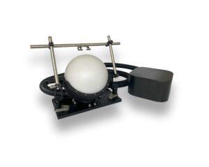
Video-based eye tracking of a Human subject displaying a vestibulo ocular reflex (VOR). The acquisition was undertaken using LabeoTech’s behavioral camera with infrared illumination and the pupil tracking was performed using the www.pupillometry.it website.
The eye is moved by three pairs of extraocular muscles that perform rotations of the ocular globe within the orbit. Compared to other body parts, such as the limbs, eye movements are relatively simpler, making it easier to classify and characterize each movement.
One common classification of eye movements is based on their role in visual function. Eye movements can be categorized into two groups: gaze-stabilizing and gaze-shifting. Gaze-stabilizing movements exist to compensate for changes in the motion of the visual scene and/or the motion of the head, while gaze-shifting movements align the eyes with a given target in the visual landscape.
Gaze-stabilizing movements include the optokinetic and vestibulo-ocular reflexes(Ambrad Giovannetti and Rancz 2024). The optokinetic reflex (OKR) occurs in response to movements of large portions of the visual field, while the vestibulo-ocular reflex (VOR) is triggered by changes in head motion. Gaze-shifting movements include saccades, which are very fast movements that shift gaze from one point to another; vergence, which involves combined changes in the angle of both eyes; and pursuit, which consists of smooth movements while following a visual target (Sekar, Panouillères, and Kaski 2024).
A lot is known about these movements, and in recent decades, eye tracking techniques have been increasingly used to better understand the underlying mechanisms of the visual system as well as motor control. Additionally, a growing body of evidence associates abnormalities in eye movements with neurodegenerative disorders such as Alzheimer’s and Parkinson’s diseases, suggesting that eye tracking can be a useful screening technique for the early detection of these disorders in the human population (Sekar, Panouillères, and Kaski 2024).
How to Measure Eye Movements
There are several methods for measuring eye movements, including electro-oculography, magnetic scleral search-coils, and video-based tracking using infrared illumination. Among these, video-based eye tracking has gained popularity in neuroscience research due to the widespread availability of high-speed cameras and the low invasiveness of the technique. Today, video-based eye tracking is widely applied across various fields of research involving both humans and laboratory animals.
The Utility of Eye Tracking in Mice
Rodents, particularly mice, have increasingly been used as animal models to investigate the structure and function of the brain. Eye tracking has not only been used to assess different aspects of visual function (e.g., OKR is widely used to assess visual acuity in mice) and eye movement control per se, but it is also an invaluable tool for probing brain states under different conditions.
Eye Tracking as a Behavioral Measurement of Brain State
The neuroanatomy of eye movement control is well understood in mice. The pathways that control eye movements involve several brain structures, including the cortex, basal ganglia, superior colliculus, brainstem, and cerebellum (Stahl 2004). This, combined with the easily quantifiable range of movements, makes eye tracking a suitable tool for the behavioral characterization of different conditions, such as pharmacological treatments or animal models of neurodegenerative diseases (de Jeu and De Zeeuw 2012; Betts-Henderson et al. 2010).
Pupil Size Tracking
Another interesting feature of video-based eye tracking is the ability to measure the dynamics of pupil contraction and dilation (pupillometry). The primary reason for performing pupillometry in mice is that changes in pupil size are closely correlated with different brain states. It has been shown that pupil size changes are linked to alertness, attention, reward prediction, and even reflect distinct brain states during sleep (Yamada and Toda 2022).
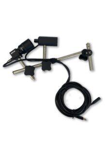
Eye tracking technique on a head-fixed mouse running on a spherical treadmill using a behavioral camera with infrared illumination. The ambient light was manipulated to induce changes in pupil size. Note the series of saccadic eye movements (~39s towards the end of the recording). The pupil tracking was performed using DeepLabCut video analysis.
Summary and Insights
Eye tracking is a powerful and versatile tool in neuroscience. Its application in murine models, particularly in the context of neurological diseases and pharmacological interventions, has proven to provide significant insights into visual function and brain states during behavior. The easy integration of eye tracking with other techniques, such as widefield optical imaging, enables researchers to extract more meaningful information from their experiments.
Products Used
References
Ambrad Giovannetti, Eleonora, and Ede Rancz. 2024. “Behind Mouse Eyes: The Function and Control of Eye Movements in Mice.” Neuroscience & Biobehavioral Reviews 161 (June):105671. https://doi.org/10.1016/j.neubiorev.2024.105671.
Betts-Henderson, Joanne, Stefano Bartesaghi, Moira Crosier, Susan Lindsay, Hai-Lan Chen, Paolo Salomoni, Irene Gottlob, and Pierluigi Nicotera. 2010. “The Nystagmus-Associated FRMD7 Gene Regulates Neuronal Outgrowth and Development.” Human Molecular Genetics 19 (2): 342–51. https://doi.org/10.1093/hmg/ddp500.
Gheorghe, Consuela-Mădălina, Victor Lorin Purcărea, and Iuliana-Raluca Gheorghe. 2023. “Using Eye-Tracking Technology in Neuromarketing.” Romanian Journal of Ophthalmology 67 (1): 2–6. https://doi.org/10.22336/rjo.2023.2.
Jeu, Marcel de, and Chris I. De Zeeuw. 2012. “Video-Oculography in Mice.” Journal of Visualized Experiments: JoVE, no. 65 (July), e3971. https://doi.org/10.3791/3971.
Jin, Xin, Suyu Chai, Jie Tang, Xianda Zhou, and Kai Wang. 2024. “Eye-Tracking in AR/VR: A Technological Review and Future Directions.” IEEE Open Journal on Immersive Displays 1:146–54. https://doi.org/10.1109/OJID.2024.3450657.
Land, Michael F. 2006. “Eye Movements and the Control of Actions in Everyday Life.” Progress in Retinal and Eye Research 25 (3): 296–324. https://doi.org/10.1016/j.preteyeres.2006.01.002.
Sekar, Akila, Muriel T N Panouillères, and Diego Kaski. 2024. “Detecting Abnormal Eye Movements in Patients with Neurodegenerative Diseases – Current Insights.” Eye and Brain 16 (April):3–16. https://doi.org/10.2147/EB.S384769.
Stahl, John S. 2004. “Using Eye Movements to Assess Brain Function in Mice.” Vision Research, The Mouse Visual System: From Photoreceptors to Cortex, 44 (28): 3401–10. https://doi.org/10.1016/j.visres.2004.09.011.
Yamada, Kota, and Koji Toda. 2022. “Pupillary Dynamics of Mice Performing a Pavlovian Delay Conditioning Task Reflect Reward-Predictive Signals.” Frontiers in Systems Neuroscience 16:1045764. https://doi.org/10.3389/fnsys.2022.1045764.
Don’t miss out! Subscribe to our newsletter for the latest blog updates.
We hope you found this post helpful! If you have any questions or need further information, feel free to reach out. We’re here to help!