Redefining the Scope of Fluorescence Imaging in Neuroscience with Shadow Imaging
Fluorescence microscopy is a key instrument in the field of neuroscience due to its capabilities in multicolor and structural imaging, coupled with advanced labeling strategies. However, certain restrictions and inconsistencies are inherent in such targeted fluorophore labelling. Enter the novel solution – shadow imaging techniques. A new approach that promises to reduce these limitations and push forward the boundaries of neuroscience research.
The Intricacies of Fluorescence Imaging
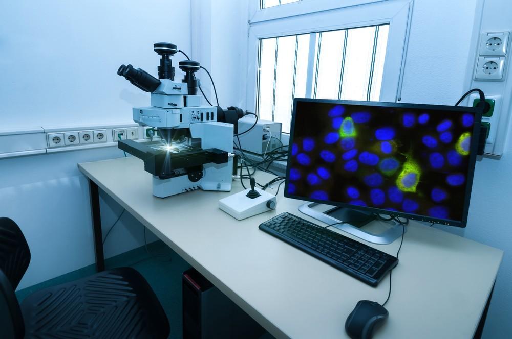
Fluorescence microscopy has become an indispensable tool in neuroscience by permitting scientists the ability to visualize neural cells with impressive specificity and diversity. This technique involves the use of fluorophore labels which attach themselves to target cellular structures. When subjected to light of a particular wavelength, these labels fluoresce, revealing complex patterns that carry significant information concerning the structure and functionality of various parts of the brain. Respectively, scientists have the ability to select fluorophores that emit light at differing wavelengths, thus separately coloring dissimilar structures in unique hues. The resulting multicolor images deliver invaluable insights into the intricate dialogues happening between the myriad of specialized cells within our brains.
Challenges with Targeted Fluorophore Labeling
Despite the crucial one-of-a-kind contributions fluorophore labeling has made in neuroscience, this method is not devoid of limitations. Firstly, it is almost impossible to achieve exhaustive or uniform labeling of targets. Unpredictable factors such as the physiological state of the sample, the penetration and retention of labels, plus their subsequent path of dissemination, can create considerable variations in label intensity within the exact same sample. Additionally, fluorescence signals can be masked or misshaped by the naturally autofluorescent factors of tissue samples. These problems complicate the process of research and could potentially skew the interpretation of results.
Shadow Imaging: A Beacon in the Shadows
Against this backdrop of challenges, comes a newly developed technique – shadow imaging – that emerges as a beacon of hope. While conventional fluorophore labeling focuses on marking the target cellular entities, shadow imaging creates an innovative approach of labeling their surroundings. The ‘shadow’ effect formed when scanned under the microscope makes the discernible outlines of the unlabeled targets visible. By not directly labeling, it eliminates the inconsistencies and inaccuracies associated with label penetration and retention. Crucially, shadow imaging can be performed on living tissue, thus opening a portal into the dynamic world of cellular structures in action. This allows neuroscientists to delve deeper into the intricacies of brain functioning, armed with real-time, detailed visualizations.
Shadow Imaging: A Supplement, Not a Substitute
Indeed, shadow imaging cannot fully replace targeted fluorophore labeling. The latter, despite its flaws, provides exceedingly precise visualizations essential for fluorescence microscopy. However, shadow imaging serves as a potent adjunct tool that significantly enhances the capabilities of fluorescence microscopy. This advanced method successfully addresses several challenges that have long been the bane of fluorescence imaging, minimizes the risk of misinterpretation, and improves the reliability of insights. The duality of comprehensive target labeling and detailed fringe context improves the quality and potential of fluorescence imaging in neuroscience.
The Future – Combining Shadow Imaging and Fluorescence Imaging
The combination of shadow imaging and fluorescence imaging represents a new frontier in neuroscience research. Both techniques offer unique advantages, but together they could provide an unprecedented perspective into the intricate architecture and functionality of the brain. By improving labeling precision and signal clarity, and enabling real-time visualization of living tissues, this synergy between shadow and fluorescence imaging could uncover new vistas of neuronal intercommunication and brain activity patterns.
As neuroscientists work to unravel the pressing complexity of the brain, innovative tools like shadow imaging emerge as invaluable aids. Though it cannot completely replace targeted fluorophore labeling, shadow imaging significantly increases the capacities of fluorescence microscopy. Powered by increased precision and added reliability, neuroscientists can now map brain structure and activity with a fresh perspective – one more step towards exploring the still vast, unknown realms of the brain.