Exploring Learning and Reward Processing with Virtual Reality in Mice
In animal research, the study of behavior has been a central part of the field of neuroscience for decades. In recent years, there has been an increasing interest in investigating the neuronal processes underlying normal and pathological brain states, such as autism, dementia and Parkinson’s disease, that impact behavior in awake animals. This has been particularly relevant for studies in rodents.
Recording brain activity during unconstrained natural behavior would be ideal to avoid experimental bias. However, this comes with several challenges. Natural behavior is rich and varied, involving multiple sensory stimuli and complex movement patterns. At the same time, this complexity can introduce variability in the data and provides limited control over sensory inputs and behavioral variables. Virtual reality has therefore appeared as an experimental alternative to fully unconstrained behavioral paradigms, offering behaviorally relevant environments while allowing stricter control over experimental variables.
Virtual Reality
Virtual reality (VR) can be described as a simulated environment in which sensory input changes in response to an individual’s actions. VR has been used in neuroscience for several decades. Initially used mainly in human research, VR later became a widely used tool for animal experiments to investigate neuronal processes involved in navigation (Attinger et al. 2025), spatial memory (Malone et al. 2024), and learning (Leinweber et al. 2017).
In mice, VR setups are generally used with head-fixed preparations, often while the animal runs on a treadmill. The head fixation allows the integration of different recording techniques such as widefield optical imaging or two-photon microscopy to record brain activity during behavioral tasks. The VR environment can be designed around a single dominant sensory modality (e.g. visual (Lee et al. 2022) or tactile (Armstrong and Vlasov 2026)) or integrate multiple ones such as auditory and visual (Cushman et al. 2013). Researchers can use VR setups to perform a variety of tasks tailored to specific features of brain function, including spatial navigation (Prince et al. 2025), task-dependent neuronal processing (Luo et al. 2025), and adapted versions of classic real-world behavioral tests such as the Y-maze (Sarpong et al. 2025), T-maze (Lee et al. 2022), Morris water maze (Cushman et al. 2013), or fear conditioning (Krishnan et al. 2025).
Experiment
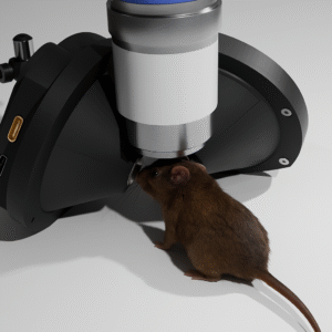
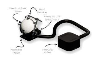
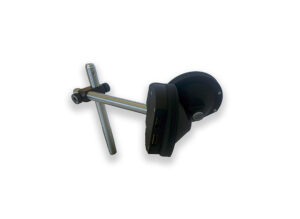
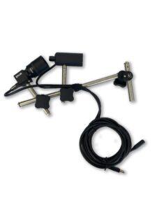
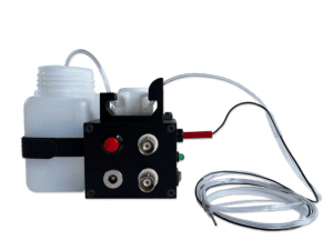
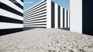
This experiment was performed with a small cohort of five mice with surgically implanted headposts. The animals were head-fixed on a spherical treadmill and the virtual reality environment was presented through a dedicated mouse VR headset. The task consisted of a 5 m linear track with walls displaying distinct visual patterns, in which the animals were trained to associate one of the wall patterns with a liquid reward (sugar water) delivered through a spout connected to a water dispenser with lickometer (figure 1). Each session consisted of 40 track traversals, and the animals performed one session per day.
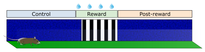
The wall patterns defined three distinct regions along the track: a control zone, a reward zone, and a post-reward zone. The control and post-reward zones consisted of blue walls with random noise and were each 2 m long. The reward zone consisted of vertical gratings and was 1 m long. The reward zone began 0.8 m before the grating-patterned wall and extended to the end of the wall.
Training Protocol
The animals were first habituated to the treadmill and the VR goggles for 5–7 days prior to this behavioral task. Once all mice were habituated to the setup, the task training was undertaken as follows:
- Days 1–3 (pre-training): the reward was automatically delivered once the animal reached the reward zone.
- Days 4–9 (training): the reward delivery was triggered by licks detected by the water dispenser device only when the animal was inside the reward zone.
Screen capture of the virtual Reality task with a side view of the animal using a behavioral camera under infrared illumination. The track is shown as the mouse perspective. Blue frames (only for the experimenter preview) indicate the frontier between zones.
Data Analysis
The VR software recorded all detected licks as well as the reward timestamps and the animal’s coordinates in the virtual environment. This information was used to calculate the lick rate, defined as the number of licks divided by the time spent in 5 cm bins of track. An association between the visual cue (i.e. vertical grating walls) and reward delivery was inferred from systematic increases in pre-reward licking, consistent with anticipatory licking behavior.
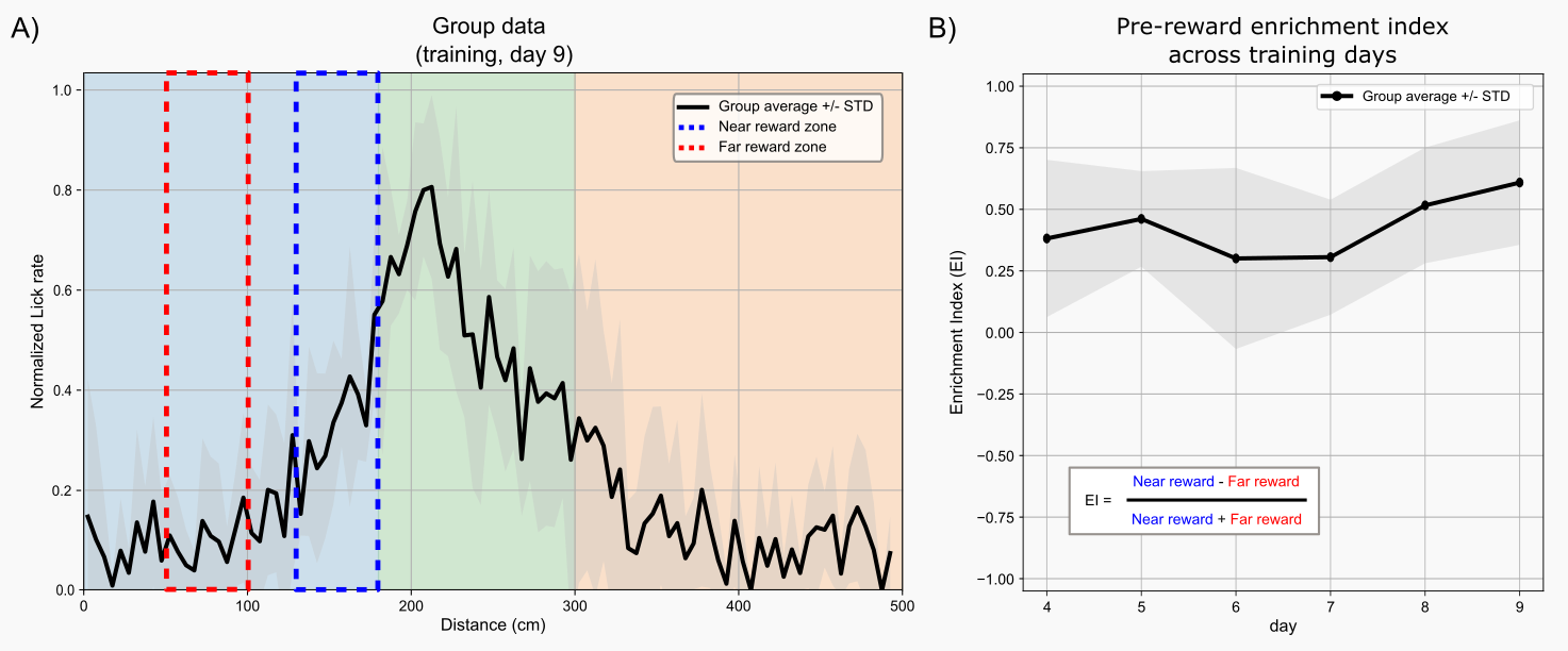
For the group analysis, the lick rate of each trial was normalized by dividing by the maximum value. A pre-reward enrichment index was calculated using a normalized difference between the average lick rate of a 50 cm window immediately before the reward zone and a baseline window (150 to 100 cm from the reward zone). Positive enrichment index values indicate an increase in licking immediately before the reward zone. The enrichment index was used as a measure of anticipatory licking behavior.
Results
All mice in the cohort developed anticipatory licking behavior associated with the reward zone (walls with vertical gratings), with some animals showing this behavior as early as day 3.
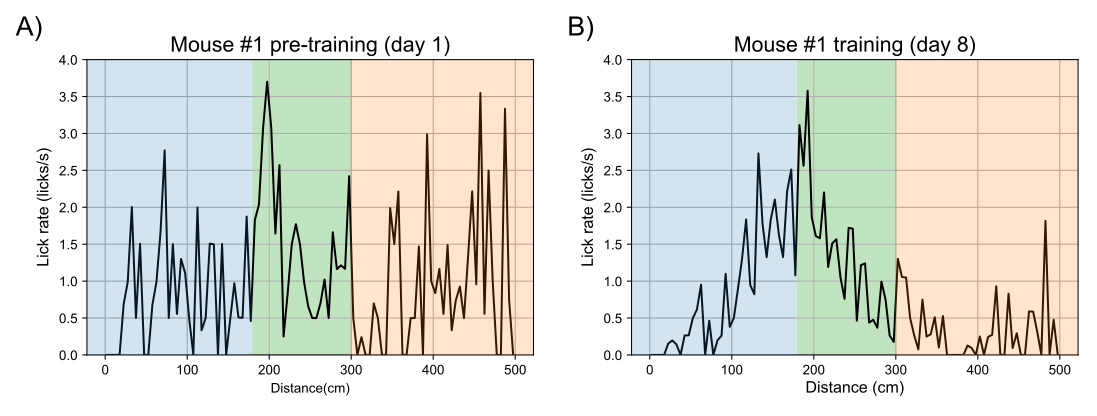
Figure 2 shows the licking pattern of a mouse on day 1 (pre-training) and day 8 (training). On day 1, the animal displayed a uniform exploratory licking pattern throughout the track while on day 8, the animal increased licking as it approached the reward zone and gradually returned toward baseline levels in the post-reward zone. This pattern was observed across all animals in the cohort at different times of the training protocol but tended to consolidate towards the end of the training period (see figure 3).
Summary and Insights
This experiment demonstrates that mice can rapidly learn to associate a visual cue with reward delivery and develop systematic anticipatory licking behavior, indicating that VR can effectively support learning and goal-directed behavior in rodents.
Virtual reality is a versatile tool for neuroscience research because it allows greater control over sensory stimuli and behavioral conditions while keeping the animal behaviorally engaged. Combined with recording techniques such as optical imaging, VR provides new possibilities to investigate the neuronal mechanisms underlying both normal and pathological brain states.
Products Used
References
Armstrong, Alex G., and Yurii Vlasov. 2026. “Neural Correlates of Perceptual Decision-Making in the Primary Somatosensory Cortex.” Proceedings of the National Academy of Sciences of the United States of America 123 (18): e2514107123. https://doi.org/10.1073/pnas.2514107123.
Attinger, Alexander, Antonia Drinnenberg, Can Dong, et al. 2025. “Environmental Novelty Modulates Rapid Cortical Plasticity During Navigation.” bioRxiv: The Preprint Server for Biology, October 22, 2025.10.21.683723. https://doi.org/10.1101/2025.10.21.683723.
Cushman, Jesse D., Daniel B. Aharoni, Bernard Willers, et al. 2013. “Multisensory Control of Multimodal Behavior: Do the Legs Know What the Tongue Is Doing?” PLOS ONE 8 (11): e80465. https://doi.org/10.1371/journal.pone.0080465.
Krishnan, Seetha, Can Dong, Heather Ratigan, Denisse Morales-Rodriguez, Chery Cherian, and Mark Sheffield. 2025. “A Contextual Fear Conditioning Paradigm in Head-Fixed Mice Exploring Virtual Reality.” eLife 14 (June): RP105422. https://doi.org/10.7554/eLife.105422.
Lee, Julie J., Michael Krumin, Kenneth D. Harris, and Matteo Carandini. 2022. “Task Specificity in Mouse Parietal Cortex.” Neuron 110 (18): 2961-2969.e5. https://doi.org/10.1016/j.neuron.2022.07.017.
Leinweber, Marcus, Daniel R. Ward, Jan M. Sobczak, Alexander Attinger, and Georg B. Keller. 2017. “A Sensorimotor Circuit in Mouse Cortex for Visual Flow Predictions.” Neuron 95 (6): 1420-1432.e5. https://doi.org/10.1016/j.neuron.2017.08.036.
Luo, Jiaqi Keith, Junhua Tan, Erin Myhre, Peter Salvino, Amy Hark, and Lucas Pinto. 2025. “A Cholinergic Mechanism Orchestrating Task-Dependent Computation across the Cortex.” bioRxiv: The Preprint Server for Biology, November 26, 2025.11.26.690825. https://doi.org/10.1101/2025.11.26.690825.
Malone, Taylor J., Nai-Wen Tien, Yan Ma, et al. 2024. “A Consistent Map in the Medial Entorhinal Cortex Supports Spatial Memory.” Nature Communications 15 (1): 1457. https://doi.org/10.1038/s41467-024-45853-4.
Prince, Stephanie M., Sarah Danielle Cushing, Teema A. Yassine, Navya Katragadda, Tyler C. Roberts, and Annabelle C. Singer. 2025. “New Information Triggers Prospective Codes to Adapt for Flexible Navigation.” Nature Communications 16 (1): 4822. https://doi.org/10.1038/s41467-025-60122-8.
Sarpong, Gideon A., Rachel Pass, Kavinda Liyanagama, et al. 2025. “Spatially Heterogeneous Acetylcholine Dynamics in the Striatum Promote Behavioral Flexibility.” Nature Communications 16 (1): 10877. https://doi.org/10.1038/s41467-025-66826-1.
Drawings used:
https://doi.org/10.5281/zenodo.3925975
https://doi.org/10.5281/zenodo. 3925935
Don’t miss out! Subscribe to our newsletter for the latest blog updates.
We hope you found this post helpful! If you have any questions or need further information, feel free to reach out. We’re here to help!